|
© Borgis - Postępy Nauk Medycznych 8, s. 577-580
*Małgorzata Kołodziejczak1, Anna Nasierowska-Guttmejer2, 3, Iwona Sudoł-Szopińska1, 4, Anna Wiączek1
Guz Buschke-Lowensteina – trudny problem interdyscyplinarny
Buschke-Loewenstein tumor – a difficult interdisciplinary problem
1Department of General Surgery, Proctology Unit, Solec Hospital, Warsaw
Head of Department: Jacek Bierca, MD, PhD Head of Unit: Małgorzata Kołodziejczak, MD, PhD 2Pathology Department, Central Clinical, Hospital of Ministry of Interior and Administration, Warsaw Head of Department: prof. Anna Nasierowska-Guttmejer, MD, PhD 3Faculty of Health Sciences, Department of Pathology, Jan Kochanowski University, Kielce Head of Faculty: prof. Stanisław Głuszek, MD, PhD 4Department of Radiology, Institute of Rheumatology, Warsaw Head of Department: prof. Iwona Sudoł-Szopińska, MD, PhD 5Department of Diagnostic Imaging, Second Faculty of Medicine, Medical University, Warsaw Head of Department: prof. Wiesław Jakubowski, MD, PhD Streszczenie
Kłykciny olbrzymie, czyli guz Buschke-Loewensteina jest chorobą spowodowaną infekcją wirusem HPV typu 6 i 11, rzadziej 16 i 18, 54 i wg klasyfikacji WHO zaliczany jest do nowotworów brzegu odbytu. Czynnikami ryzyka zachorowania jest zakażenie wirusem HIV, zachowania homoseksualne, immunosupresja oraz choroby obniżające odporność, takie jak cukrzyca czy przewlekły alkoholizm Rozpoznanie opiera się na wywiadzie, badaniu klinicznym, histologicznym, badaniach endoskopowych i badaniach obrazowych. Obraz kliniczny jest bardzo charakterystyczny. Guz ma duże rozmiary (powyżej 10 cm średnicy). W większości przypadków rośnie powoli, przez parę lat, rzadko daje przerzuty wykazując miejscową złośliwość. Może naciekać okoliczne struktury anatomiczne, może też wtórnie powodować przetoki i ropnie. Diagnostyka przedoperacyjna powinna obejmować test na wirus HIV, ultrasonografię transrektalną lub/i MR, która wykazuje stosunek masy guza do mięśni zwieraczy odbytu, CT miednicy, w przypadku naciekania guza na narządy sąsiednie oraz kolonoskopię. Najczęściej stosowaną metodą leczenia jest miejscowe wycięcie guza. Jeżeli występuje naciekanie guza na zwieracze odbytu lub naciekanie guza na sąsiednie narządy – konieczne jest wyprowadzenie stomii, a czasem brzuszno-kroczowe odjęcie odbytnicy. Chemioterapia ma znaczenie drugoplanowe, a skuteczność radioterapii jest dyskusyjna. Konieczne są dalsze multidyscyplinarne badania celem dokładniejszego poznania choroby i określenia skutecznych sposobów leczenia. Słowa kluczowe: kłykciny olbrzymie, guz Buschke-Loewensteina, HPV
Summary
Giant condylomaacuminatum (also known as a Buschke-Lowenstein tumor and giant condyloma of Buschke-Lowenstein tumor) is a disease caused by an infection with HPV type 6 and 11, rarely 16, 18, 54 and according to the WHO, is classified as one of the anal verge neoplasms. Risk factors for the disease are: an infection with HIV, homosexuality, immunosupression and diseases which lower immune system, such as diabetes and chronic alcoholism. Diagnosis is based on clinical examination, histopatological assessment, endoscopic and imaging studies. Clinical presentation is very characteristic. The tumor is large (above 10 cm in diameter). In most cases it is a slow-growing, even up to several years, lesion, which rarely metastases, is locally destructive, may invade surrounding structures, and can also lead to the formation of anal fistulas and abscesses. Preoperative diagnosis should include: HIV testing, anorectalendosonography or MRI, which presents relation of the tumor to anal sphincters, as well as the pelvis, CT, in case of tumor infiltration into the surrounding organs, colonoscopy. The most common method of treatment is a local surgical excission of the tumor. If an infiltration of the tumor to the anal sphincter or tumor invasion into adjacent organs is present – it is necessary to perform a colostomy and sometimes, an abdominoperineal excision of the anus. Chemotherapy is of secondary importance and the effectiveness of radiotherapy is questionable. Multi-disciplinary studies need to be performed to learn more about the disease and to identify effective methods of treatment. Key words: Giant condylomaacuminatum, Buschke-Lowenstein tumor, HPV
Introduction
Buschke-Lowenstein tumor (BLT) termed the genital giant (giant condyloma), according to the classification of the World Health Organization is a variant of squamous cell carcinoma of the anal canal bank called papillary carcinoma (verrucous carcinoma) (tab. 1) (1). It macroscopically resembles warts (condylomata acuminata), but is larger in size. The rate of BLT in a normal population is low, at 0.1%, while it increases in the male homosexual population. Warts are transmitted sexually and develop as a result of infection with human papillomavirus (HPV). Anal verge cancers are morphologically similar to other tumors of the skin, but are less aggressive than the tumors of anal canal, and more of them vary in terms of histopathology. The incidence of anal verge cancer is about five times smaller than the tumors of the anal canal (2). In most cases patients with Buschke-Lowenstein tumor HPV infection is detected, mainly types 6 and 11, 16 and 18 less frequently and very rarely 54. This may be helpful in differentiating with cancer (squamus cell carcinoma), where mainly virus types 16 and 18 is detected. Currently, over 120 HPV types have been distinguished, which generally can be divided into high risk HPV (e.g., types 16, 18, 33, 34, 45, 52, 56) and low risk HPV (e.g., types 6, 11, 42, 43, 44, 55). Interestingly, the most common ones in patients with HIV infection are warts caused by benign HPV 6 and HPV 11, which are also detected in the Buschke-Loewenstein tumor. The tendency to malignant transformation in Buschke-Loewenstein tumor is much higher than in the case of “typical” warts, approximately 56% (3). The average time of malignant transformation is 5 years (mean 15-100 months) (3).
Table 1. Histological classification of tumors of the anus (21).
The clinical form of giant condyloma was first described by Buschke and Loewenstein in 1925 (4). The authors present the case of a patient with giant warts located on the penis, describing the change as “invasively growing warts-like tumor”. The perianal location of Buschke-Loewenstein tumor was first described by Peiron in 1931, a 48-year-old patient. Buschke-Loewenstein tumor is more common in men than in women, usually under 50 years of age (the mean 43 years). Trombetta et al. (5) on the basis of the review of scientific literature from the years 1958 to 2000 identified 51 cases of Buschke-Loewenstein tumor. Men got sick more often (2.7:1), the mean age of patients was 43.9 years. Isolated cases of this tumor are also described in children (6). Ambriz-González et al. (6) presented the case of a 12-year-old girl with a Buschke-Loewenstein tumor in the anal area treated effectively with local excision and radiotherapy. Risk factors for developing giant warts are infections with HIV, homosexuality, immunosuppression (e.g. transplant patients after surgery) and immunosuppressive diseases, such as diabetes or chronic alcoholism. In the case of HIV-positive men 92% of HPV infection is recognized around the anus.
Diagnosis is based on history, clinical examination, histological examination, and endoscopic and imaging examinations (transrectal ultrasound, MRI, CT).
Clinical picture is very distinctive. The tumor is large (over 10 cm in diameter) (fig. 1). In most cases, grows slowly, for a few years, rarely metastasizes, exhibits local malice. It may invade surrounding anatomical structures (vagina, urethra, prostate, anal sphincters), it can also cause secondary fistula and abscess. It occasionally invades retroperitoneal space above the anal lifters muscles. Soós et al. (7) described a 63-year-old woman with a Buschke-Loewenstein tumor located on the edge of the anus. The tumor infiltrated the anal sphincter muscle and superalevator retroperitoneal space, causing bilateral hydronephrosis. Patients with a Buschke-Loewenstein tumor may report pain, bleeding, itching and burning of inflamed skin around the anus, and in the case of invasion of the tumor to the anal sphincter, urinary and bowel gas. On the surface of the tumor abscess or anal fistula may occur. According to Trombetta et al. (5) pain in the course of Buschke-Loewenstein tumor is reported by 32% of patients, bleeding in 18% of cases, abscess and fistula can develop in 32% of patients.
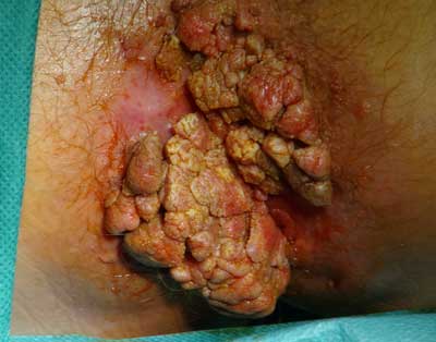 Fig. 1. The clinical picture of Buschke-Loewenstein tumor.
Histopathology
Buschke-Loewenstein tumor (verrucous carcinoma) requires differentiation based on histopathological examination of: overgrown usual classical genital warts and squamous cell carcinoma of the vulva, penis and anus. Differential diagnosis between the overgrown warts, and papillary carcinoma is difficult.
The essential macro- and microscopic features of these changes are the size and how they grow. Common warts’ histopathology is characterized by hyperplasia and superficial squamous epithelium forming papillary structures. BLT (papillary carcinoma) develops as large pendula lumpy mass, usually more than 12 cm in diameter, composed of an exophytic and endophytic parts with characteristic dilating and oppressing stroma margin (pusching margin). Classical squamous cell carcinoma forms an invasive tumor with the presence of irregular scattered tumor nests infiltrating carcinoma fibrous stroma. Microscopically it creates a distinctive image of acanthotic and expanding papillary squamous epithelium with preserved stratification. Inside the tumor it forms galls invading stroma with tongues, and, in places, cystic masses filled with hyperkeratotic structures (horn-like) (8). Epithelial cells look like in a mild tumor, they have a large nucleus with nucleolus, a small polymorphism and distribution figures restricted to basal layers. BLT malignancy is intermediate between the warts and classic squamous cell carcinoma. Papillary carcinomas are a rare variant of well-differentiated squamous cell carcinoma SCC, a mild one, with a tendency to relapse and a low risk of metastasis (9). Poor prognosis is associated with the polymorphism of stromal cells and infiltrating stroma by scattered cells lying in small nests or being dispersed. Such cases should be diagnosed and treated as classical squamous cell carcinoma.
If the location of the Buschke-Loewenstein tumor is the sole of the foot a change takes place to an exophytic character with fissures and bays, filled with horn-like masses, resembling branched rabbit burrows, hence the Latin name carcinoma (epithelioma) cuniculatum, meaning a rabbit burrow (9).
Accurate diagnosis determines the selection of an appropriate therapeutic approach (10).
The preoperative also includes performance:
– a test for HIV,
– IMAGING (transrectal ultrasound, MRI or CT),
– colonoscopy (which is not always technically possible due to the size of the tumor).
Transrectal ultrasound and MRI allow precise determination of the ratio of the tumor (infiltration) to the anal sphincters (internal and external) and the ani levator, an assessment of its echostructure, vascularization, possible infiltration of the adjacent structures. In the case of tumors of considerable dimensions, resulting in the stenosis of the anal canal, there is no possibility of introducing an ultrasound head to the anus. The alternative is a pelvic MRI or CT, which are the methods of choice for the assessment of tumor infiltration into adjacent pelvic organs.
Colonoscopy is a supplement of relevant imaging.
Treatment
Due to the size of the tumor, treatment is difficult. The most common method is its local resection (11). In the available literature, the success rate using this method reaches 45.5%. In the Department of Proctology in the Solec Hospital a several stages resection is used, sometimes combined with covering the loss with a cutaneous flap. If possible, light radial cuts of the anal canal are applied. When surgical cutting the Buschke-Loewenstein tumor one should observe the following rules:
– if the tumor circumferentially covers the entire circumference of the anus, keep healthy anodermis bridges between the wounds (or even warts to be removed in the 2nd stage) to make the extensive wound healing possible,
– wounds should not be stapled close, but left partially open to drain the circuit,
– when multi-stage resection is used, subsequent treatments are to be carried out at intervals of about 4 weeks.
In the case of tumor infiltrating anal sphincters or adjacent organs, it is necessary to have the stoma resection, and sometimes abdominoperineal rectum resection is needed (12). The prophylactic inguinal lymph nodes are not prophylactically resected due to the low probability of metastasis. In the literature, there are also numerous reports concerning the healing of the Buschke-Loewenstein tumor after the application of a wide resection of local changes with S-plasty without performing colostomy (13). Some surgeons combine classic resection of warts with CO2 laser therapy (14), while others obtain good results of the concomitant of chemoradiotherapy with surgery (15). It should be mentioned that medical personnel infections with viral particles present in the smoke during the laser treatment are described (16). In the case of a patient infected with HIV chemotherapy is a problem, it can in fact activate immune disorders. Generally, chemotherapy is used in the case of tumor recurrence. Opinions on the effectiveness of radiotherapy in the treatment of Buschke-Loewenstein tumor are divided (17). Sobrado et al. (18) show good results using telecobalt therapy. Antiviral drugs in the form of acting locally cidofovir 1.5% or interferon can be used only after the resection of changes. Similarly, imiquimod, acting rather locally, is used in the prevention of relapses. For the prophylaxis of recurrences autologous vaccines are also recommended. In the literature, one case of spontaneous remission of the tumor in a pregnant patient after giving birth by caesarean section is described (19). After resection regular checks are necessary because of the high recurrence and possible malignant metaplasia (20).
Complications of the disease result from the large size of the tumor, which invades the surrounding tissue. These include:
– inflammation of the skin and subcutaneous tissue,
– abscesses and fistulas,
– anal canal stenosis,
– incontinence (in the case of infiltration of the sphincter),
– bleeding from the tumor.
Complications associated with the surgery are:
– bleeding from the wound,
– skin defects and poor cosmetic results,
– narrowing tissue scars in the anal canal.
Recurrence following surgery occurs in 61% of cases. Recurrences are more frequent in HIV-positive patients and are often associated with reduced levels of CD4 (immunosuppressed). Mortality from Buschke-Loewenstein reaches tumor is 21% of cases.
Conclusions
1. Buschke-Loewenstein tumor is a tumor of local malignancy because of the large size and infiltration of surrounding tissue as an expansion.
2. In a patient with giant warts HIV infection should always be ruled out.
3. Wide radical resection, often with several stages is the treatment of choice, leaving the wound to heal per secunda or using reconstructive techniques (moved myocutaneous flap). It often requires the removal of temporary stoma, in the case of recurrence – performing abdominoperineal rectum resection.
4. Chemotherapy is of secondary importance.
5. Adjuvant therapies, such as radiation therapy and immunotherapy may bring good benefits, but their effect is still not fully understood.
6. More multi-disciplinary research is needed in order to fully understand the pathogenesis of the disease and identify effective ways of treatment. Piśmiennictwo
1. Bosman FT, Carneiro F, Hruban RH, Thiese ND (eds.): WHO Classification of Tumour of the Digestive System. IARC, Lyon 2010: 183-185.
2. Mik M, Dziki A: Nowotwory odbytu. [W:] Dąbrowski A (red.): Gastroenterologia. Cz. II, Medical Tribune 2011: 398-401.
3. Chu QD, Vezeridis MP, Libbey NP, Wanebo HJ: Giant condyloma acuminatum (Buschke-Loewenstein tumor) of the anorectal and perianal regions. Analysis of 42 cases. Dis Colon Rectum 1994; 37: 950-957.
4. Buschke A, Loewenstein L: Über carcinomahnliche condylomata acuminata des penis. Klin Wochenschr 1925; 4: 1726-1728.
5. Trombetta LJ, Place RJ: Giant condyloma acuminatum of the anorectum: trends in epidemiology and management. Report of a case and review of the literature. Dis Colon Rectum 2001; 44: 1878-1886.
6. Ambriz-González G, Escobedo-Zavala LC, Carrillo de la Mora F et al.: Buschke-Löwenstein tumor in childhood: a case report. J Ped Surg 2005 Sep; 40(9): 25-27.
7. Soós Z, Varga T, Vadinszky P et al.: Verrucous carcinoma of the anal margin. The importance of adequate biopsy technique. Orv Hetil 2011 Feb 27; 152(9): 344-348.
8. Mazurkiewicz W: Choroby przenoszone drogą płciową. [W:] Bielecki K, Dziki A (red.): Proktologia. PZWL, Warszawa 2000: 313-336.
9. Bieniek A, Cisło M, Matusiak Ł et al.: Rak brodawkujący (carcinoma verrucosum) – przegląd objawów klinicznych i histologicznych. Post Dermatol Alergol 2006; XXIII(2): 57-66.
10. Longacre TA, Kong CS, Welton ML: Diagnostic problems in anal pathology. Adv Anat Pathol 2008 Sep; 15(5): 263-278.
11. Renzi A, Giordano P, Renzi G et al.: Buschke-Lowenstein tumor successful treatment by surgical excision alone: a case report. Surg Innov 2006 Mar; 13(1): 69-72.
12. Papiu HS, Dumnici A, Olariu T et al.: Perianal giant condyloma acuminatum (Buschke-Löwenstein tumor). Case report and review of the literature. Chirurgia (Bucur) 2011 Jul-Aug; 106(4): 535-539.
13. De Toma G, Cavallaro G, Bitonti A et al.: Surgical management of perianal giant condyloma acuminatum: Report of three cases. Eur Surg Res 2006; 38: 418-422.
14. Martin JM, Molina I, Monteagudo C et al.: Buschke-Lowenstein tumor. J Dermatol Case Rep 2008 Dec 27; 2(4): 60-62.
15. Tytherleigh MG, Birtle AJ, Cohen CE et al.: Combined surgery and chemoradiation as a treatment for the Buschke-Löwenstein tumour. Surgeon 2006 Dec; 4(6): 378-383.
16. Grochowicz M, Grochowicz P: Kłykciny kończyste. [W:] Bielecki K, Dziki A (red.): Proktologia. PZWL, Warszawa 2000: 313-336.
17. Mudrikowa T, Jaspers C, Ellerbroek P, Hoepelman A: HPV- -related anogenital disease and HIV infection: not always “ordinary” condylomata acuminate. J Med 2008; 66(3): 98-102.
18. Sobrado CW, Mester M, Nadalin W et al.: Radiation-induced total regression of a highly recurrent giant perianal condyloma: report of case. Dis Colon Rectum 2000 Feb; 43(2): 257-260.
19. Picaud A, Faye A, Ogowet-Igumu N et al.: Büschke-Loewenstein tumor during pregnancy. Apropos of 2 cases. Rev Fr Gynecol Obstet 1990 Jun; 85(6): 375-378.
20. Lévy A, Lebbe C: Buschke-Löwenstein tumour: diagnosis and treatment. Ann Urol (Paris) 2006 Jun; 40(3): 175-178.
21. Fenger CF, Frisch M, Marti MC, Parc R: Tumours of anal canal. [In:] Hamilton SR, Aaltonen LA: World Health Organization of Tumours: Pathology and Genetics of Tumours of the Digestive system. IARC Press, Lyon 2000: 145-155.
otrzymano/received: 2013-05-15 zaakceptowano/accepted: 2013-06-26 Adres/address: *Małgorzata Kołodziejczak Department of General Surgery, Proctology Unit Solec Hospital ul. Solec 93, 00-382 Warszawa tel.: +48 (22) 250-62-68 e-mail: drkolodziejczak@o2.pl Artykuł Guz Buschke-Lowensteina – trudny problem interdyscyplinarny w Czytelni Medycznej Borgis. |
  Proszę kliknąć w wybraną okładkę aby przejść na stronę czasopisma
 |


Chcesz być na bieżąco? Polub nas na Facebooku: strona Wydawnictwa na Facebooku |