|
© Borgis - Postępy Nauk Medycznych 10, s. 758-764
*Anna Lis-Święty1, Joanna Gola2, Urszula Mazurek2, Ligia Brzezińska-Wcisło1
Ekspresja genów kodujących transformujący czynnik wzrostowy β1 i jego receptory u chorych z twardziną układową i objawem Raynauda
Transforming growth factor β1 and its receptors gene expression in patients with systemic sclerosis and Raynaud’s phenomenon
1Dermatology Department, Medical University of Silesia, Katowice
Head of Department: prof. Ligia Brzezińska-Wcisło, MD, PhD 2Department of Molecular Biology, Medical University of Silesia, Sosnowiec Head of Department: prof. Urszula Mazurek, MD, PhD Streszczenie
Wprowadzenie. Rolę TGFβ1 w patogenezie twardziny układowej (ang. systemic sclerosis – SSc) wykazano we wcześniejszych badaniach dotyczących ekspresji tego czynnika w tkankach. Cel pracy. Celem pracy było zbadanie jak się zmienia liczba kopii mRNA genów kodujących TGFβ1 i jego receptory w leukocytach krwi obwodowej chorych z SSc i objawem Raynauda (ang. Raynaud’s phenomenon – RP). Materiał i metody. Badaniem objęto 19 chorych z SSc, 8 pacjentek z RP i 8 osób zdrowych stanowiących grupę kontrolną. Oznaczenie mRNA genów kodujących TGFβ1, TGFβRI, TGFβRII oraz TGFβRIII przeprowadzono techniką ilościowej reakcji amplifikacji z odwrotną transkrypcją w czasie rzeczywistym (ang. Real-Time Quantitative Reverse Transcription Polymerase Chain Reaction, real-time QRT-PCR). Wyniki. Liczba kopii mRNA dla TGFβ1 była znacząco wyższa u chorych z SSc w porównaniu z grupą chorych z RP. W obu grupach ekspresja mRNA dla TGFβ1 była znacząco wyższa niż w grupie kontrolnej. Nie stwierdzono znaczących różnic w ekspresji mRNA genów kodujących receptory TGFβ1 pomiędzy grupą SSc a pacjentami z RP. Stosunki TGFβ1/TGFβRI mRNA i TGFβRIII/TGFβRI mRNA były znacząco wyższe u chorych z SSc w porównaniu z grupą RP. W grupie chorych z SSc i kontrolnej liczba kopii mRNA dla TGFβ1 korelowała z liczbą kopii mRNA dla TGFβRIII. W grupie kontrolnej mRNA dla TGFβRI i mRNA dla TGFβRII korelowały dodatnio (w obu rosły), podczas gdy w obu grupach chorych korelacja ta była negatywna, co oznacza, że jeżeli jeden parametr rośnie, to drugi maleje. Wnioski. Dysregulacja ekspresji genów kodujących TGFβ1 i jego receptory w SSc i RP może przekładać się na zmianę aktywności TGFβ1, co może pociągać za sobą zapoczątkowanie procesu zapalnego, a następnie włóknienia. Słowa kluczowe: Real-time QRT-PCR, TGFβ1, twardzina układowa, objaw Raynauda
Summary
Introduction. Previous studies concerning the tissue expression of TGFβ1 demonstrated that this factor may play the role in the pathogenesis of systemic sclerosis (SSc). Aim. To examine the change in the number of mRNA copies of genes coding TGFβ1 and its receptors in peripheral blood leucocytes in patients with SSc and RP. Material and methods. The research concerned 19 patients with SSc, 8 patients with RP and 8 healthy persons constituting the control group. Quantification of TGFβ1, TGFβRI, TGFβRII, TGFβRIII genes mRNA was carried out with the use of Quantitative Real-Time Reverse Transcription Polymerase Chain Reaction. Results. The number of copies of TGFβ1 mRNA was significantly higher in patients with SSc than in the group of patients with RP. In both groups the TGFβ1 mRNA level was significantly lower than in control group. No significant differences were found between SSc and RP patients when mRNAs of genes coding TGFβ1 receptors were analyzed. The TGFβ1/TGFβRI mRNA and the TGFβRIII/TGFβRI mRNA ratios were significantly higher in patients with SSc than in RP patients. In the group of patients with systemic sclerosis and in control group the number of copies of TGFβ1 mRNA correlated with the number of copies of TGFβRIII mRNA. In control group TGFβRI mRNA and TGFβRII mRNA correlated positively (both of them were increasing), while in both groups of patients this correlation was negative, what means that one parameter was increasing when the second one was decreasing. Conclusions. Disregulation TGFβ1 and its receptors gene expression in SSc and RP may translate to changes in the activity of TGFβ1 which may result in the initiation of the inflammatory process and later fibrosis. Key words: Real-time QRT-PCR, TGFβ1, Systemic sclerosis, Raynaud phenomenon
Introduction
Systemic sclerosis (SSc) is a disease of the connective tissue characterized by vascular changes and immunological dysfunctions which lead to progressive skin and internal organ fibrosis. First clinical manifestation is usually Raynaud’s phenomenon (RP) (paroxysmal blanching with subsequent cyanosis and swelling of hands) connected with a generalized vasculopathy of minor vessels in the skin and internal organ areas (1). A consequence of rupturing the endothelium is a migration of mononuclear peripheral blood cells to the extravascular space and creation of inflammatory infiltrations characteristic for SSc (2, 3). T-cells and monocytes are dominant in this infiltration. Whilst producing a series of cytokines and growth factors these cells are able to initiate a series of intercellular interactions leading to vessel changes as well as disregulation of synthesis and degradation of extracellular matrix components. The main role in the fibrosis processes in SSc could be played by transforming growth factor β1 (TGFβ1) produced in excess by peripheral blood mononuclear cells (PBMC) (4). It has been proved that this cytokine can stimulate gene transcription of collagen by stimulation of synthesis or activation of specific transactive DNA binding factors (5, 6). In patients with limited SSc (lSSc) and diffuse SSc (dSSc) treated with pamidronate (aminobisphosphonate) a approx. 30% decrease in TGFβ1 production by the PBMC was noticed which could explain a positive therapeutic effect (7). Hasegawa et al. (8) demonstrated an increase in TGFβ1 production by PBMC in patients with SSc in comparison with a healthy persons control group. However, this data was not confirmed in other papers. In research of Giacomelli et al. (9) the TGFβ1 concentration in the serum and supernatants of the PBMC culture from SSc patients in spontaneous conditions as well as after phytohemagglutinin (PHA) stimulation was not different from the control group. The concentration of TGFβ1 in serums of patients with SSc can remain unchanged, reduced in relation to the control group or be below the lower limit of detection, however TGFβ1 can be present in large amounts in the tissue (10-14). Reasons for this are faintly sensitive methods or inhibitors appearing in the serum. In physiologic states TGFβ1 binds with proteins (latency-associated peptide, α2-macroglobulin), which can largely conceal its presence in the blood and are responsible for a non-linear diagnosed sample dilution curve line and a divergence in relation to the standard curve in the ELISA method (15, 16).
An analysis of gene expression in the earlier stage of this process, that is the transcription level, not only doesn’t possess such limitations but also allows for the detection of molecular changes preceding changes at protein level. Therefore the main aim of the study was to evaluate the number of mRNA copies of genes coding TGFB1 and its receptors in peripheral blood leukocytes changes in patients with SSc and isolated RP in which capillaroscopy and (or) immunological markers presence suggested a risk of SSc development.
material and methods
The study group consisted of 27 patients (26 women and 1 man) with RP, aged 18 to 65, average 48.1 ± 11.6 years hospitalized in Medical University of Silesia – Dermatology Department in Katowice with a suspicion or diagnosed SSc. The RP lasted for 0.3 to 25 years, average 8.2 ± 5.9 years. A capillaroscopy examination of the nailfold was carried out for each patient, antinuclear antibodies were marked with the indirect immunofluorescence (IIF) method on Hep-2 cells and detailed diagnostic research was conducted allowing for an assessment of the internal organs affected by pathological changes. Changes in the esophagus were diagnosed based on confirmed peristaltic dysfunction and/or smoothing out the mucous membrane folds in a radiological examination of the esophagus. Influence on the lungs was attested to bilateral fibrosis changes in chest X-ray. Cardiologic changes with characteristics of arrhythmia, conductivity disorders in ECG examination or during transesophageal electrostimulation and syndromes of right ventricle failure, prior to lung hypertension were diagnosed as heart muscle involvement in course of SSc. Influence on the kidney by the disease process was diagnosed based on a persistent proteinuria and coexisting arterial hypertension. Myositis type muscle changes aside from clinical symptoms: muscle weakness and pain were diagnosed based on increased activation of muscle enzymes (creatine phosphokinase and aldolase) as well as aberrations in electromyography and histopathology examinations. Apart from this, routine laboratory tests were carried out: ESR, blood morphology, general urine test. Other tests were: Waaler-Rose reaction, latex-R, electrophoresis of serum protein division and assessment of kidney functions.
In 19 patients SSc was diagnosed based on the American College of Rheumatology (17) criteria, remaining 8 were female patients with an isolated RP. Skin changes in patients with SSc corresponded with lSSc – appeared on the skin of the face, upper limbs up to 1/3 of the forearm. Table 1 presents a clinical characteristic of patients with RP without clinical symptomes of connective tissue diseases and patients with lSSc. Patients qualified for the research were not treated earlier with immunosuppressive agents and (or) steroids. Control samples were obtained from 8 healthy volunteers. The Medical University of Silesia Local Research Ethics Committee approved the study and all subjects provided informed consent to participate.
Table 1. Clinical characteristics of patients with isolated RP and patients with lSSc.
Extraction of total RNA
Total RNA was isolated from the 500 μl whole blood samples using acid guanidinium-thiocyanate phenol-chloroform method (18). Extracts of total RNA were purified with the use of RNeasy Mini Kit (Qiagen Gmbh, Hilden, Germany), in accordance with manufacturer protocol. The quality of RNA was estimated by electrophoresis on a 1% agarose gel stained with ethidium bromide. The RNA concentration was determined by absorbance at 260 nm using a Gene Quant II spectrophotometer (Pharmacia LKB Biochrom Ltd., Cambridge, UK).
mRNA quantification by Quantitative Real-Time Reverse Transcription Polymerase Chain Reaction
The quantitative analysis was carried out with the use of Sequence Detector ABI PRISM™ 7000 (Applied Biosystems, Kalifornia, USA). The quantity of PCR products was determined after each round of amplification, using fluorescent dye SYBR Green I (Qiagen Gmbh, Hilden, Germany) that binds double-stranded DNA. The standard curve was appointed for standards of β-actin (Applied Biosystems, Kalifornia, USA). For this assay positive (β-actin mRNA) and negative (no template) controls were carried out. The nucleotide sequences of the PCR primers used to assay gene TGFβ1, TGFβR1, TGFβR2, TGFβR3 and β-actin (endogenous control) expression, chemical and thermal conditions of amplification were as previously (19-21).
Sequence specificity of amplimers
Sequence specificity of amplimers was proved by analysis with ABI PRISM™ 377 DNA Sequencer (Applied Biosystems, Kalifornia, USA). Melting temperatures of amplimers were assessed by SYBR Green I Dissociation assay (Dissociation Curve Software – Applied Biosystems, Kalifornia, USA). The PCR products and molecular weight marker pBR 322/Hae III (Fermentas International Inc., Ontario, Canada) were separated on 8% polyacrylamide gel and visualized using silver staining (LKB-Pharmacia). The length of amplified fragments was assessed by analysis with GelScan v.1.45 software (Kucharczyk TE, Warsaw, Poland).
Statistical analysis
The values were expressed as median and range. Quantitative data were compared by a nonparametric Mann-Whitney U test. Correlations were evaluated using the Spearman rank correlation coefficient test. P < 0.05 was considered significant. All calculations were performed with Statistica Version 6.0 software (StatSoft Inc., Oklahoma, USA). The expression of the TGFβ1, TGFβR1, TGFβR2, TGFβR3 and β-actin genes was expressed as a ratio of the mRNA copy number to the 1 μg of total RNA in samples studied.
Results
β-actin mRNA
In all samples analyzed mRNA of β-actin gene was demonstrated, thus indicating the integrity of the RNA extracts.
TGFβ1 mRNA
The number of copies of TGFβ1 mRNA was significantly higher in patients with SSc than in the group of patients with RP (p = 0.0384) (fig. 1, tab. 2). In both groups the TGFβ1 mRNA level was significantly lower than in control group (SSc: p = 0.0257; RP: p = 0.0023).
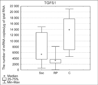 Fig. 1. Comparison of number of TGFβ1 mRNA copies in studied groups. Medians ± quartiles and extreme values of copy numbers are presented. C – control group.
Table 2. Number of mRNA copies and ratios of TGFβ1 and its receptors mRNA in studied groups.
TGFβRI, TGFβRII, TGFβRIII mRNA
No significant differences were found between SSc and RP patients when mRNAs of genes coding TGFβ1 receptors were analyzed. Only in case of TGFβRII mRNA slight trend toward difference between groups was observed, where number of copies was higher in patients with systemic sclerosis (p = 0.0844). Comparing to controls the expression of TGFβRI was significantly lower in SSc patients (p = 0.0146) (fig. 2), the number of TGFβRIII mRNA copies was significantly lower in RP patients (p = 0.0274) (fig. 3).
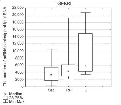 Fig. 2. Comparison of number of TGFβRI mRNA copies in studied groups. Medians ± quartiles and extreme values of copy numbers are presented. C – control group.
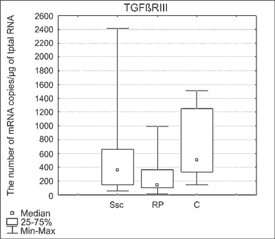 Fig. 3. Comparison of number of TGFβRIII mRNA copies in studied groups. Medians ± quartiles and extreme values of copy numbers are presented. C – control group.
The TGFβ1 and TGFβ receptors ratios
The TGFβ1/TGFβRI mRNA and the TGFβR3/TGFβR1 mRNA ratios were significantly higher in patients with SSc than in RP patients (p = 0.0079 and p = 0.0337, respectively). No significant changes were found in SSc and RP patients comparing to controls.
Correlations
In the group of patients with systemic sclerosis the number of copies of TGFβ1 mRNA correlated with the number of copies of TGFβRIII mRNA (p = 0.0004, R = 0.7281). In this group the number of copies of TGFβRI mRNA was increasing when the number of copies of TGFβRII mRNA was decreasing (p = 0.0081, R = -0.5877). Trend toward correlation between TGFβRII mRNA and TGFβRIII mRNA was also observed (p = 0.0675, R = 0.4281). In the group of patients with isolated Raynaud phenomenon only trend toward correlation between TGFβRI mRNA and TGFβRII mRNA was observed (p = 0.071, R = -0.666). Like in group of patients with systemic sclerosis the number of copies of TGFβRI mRNA was increasing when the number of copies of TGFβRII mRNA was decreasing. In the control group, like in the group of SSc patients, the number of copies of TGFβ1 mRNA correlated with the number of copies of TGFβRIII mRNA (p = 0.002, R = 0.7281). Trend toward correlation between TGFβRI mRNA and TGFβRII mRNA was also observed (p = 0.071, R = 0.6666 the same strength like in RP patients). Surprisingly, in control group TGFβRI mRNA and TGFβRII mRNA correlated positively (both of them were increasing), while in both groups of patients this correlation was negative, what means that one parameter was increasing when the second one was decreasing.
Discussion
Transforming growth factors β-TGFβ constitute the glikoprotein family of which thus far 3 factors: TGFβ1, TGFβ2 and TGFβ3 are known in people (22). Best known is TGFβ1, isolated from blood platelets about 20 years ago. This cytokine is currently considered one of the most important factors in etiopathogenesis of SSc. In regard of numerous problems connected with assaying of TGFβ1protein in serum of patients with SSc in this study we analysed transcriptional activity of TGFβ1 gene. The number of mRNA copies of genes coding TGFβ1 and its receptors was determined by Quantitative Real-Time Reverse Transcription Polymerase Chain Reaction (real-time QRT-PCR) (23) technique which has been used for several years in many branches of medicine. The material for our research was peripheral blood because no increase of TGFβ1 synthesis in fibroblasts of patients with SSc was noticed (24), however it is considered that the main sources of TGFβ1 are monocytes, T cells and also blood platelets (25) which take part in the disease pathogenesis. Our research also included patients with an isolated RP in which capillaroscopy and (or) immunological markers presence suggested a risk o SSc development. In patients with SSc number of TGFβ1 mRNA copies was significantly higher than in patients with RP. Both in the group of the patients with SSc and RP the number of mRNA copies of this gene was significantly lower than in the control group. Research up to date showed that TGFβ1 has most significance in early development stage of SSc (26-27). However data concerning the role of TGFβ1 in patients with an isolated RP are insufficient. Angiokinetic changes can precede the development of skin sclerosis by many years and are a clinical manifestation of damage to the endothelial tissue and blood platelets activation. Mortazavi-Haghighat et al. showed a significant increase in TGFβ1 expression and its receptors in vessels and skin fibroblasts in conditions of hypoxia (38). This is probably a positive phenomenon as in disorders connected with ischemia and reperfusion a protective effect of this cytokine has been shown. TGFβ1 strongly slows down the peripheral blood mononuclear adhesion to the endothelium (29) and free radicals generation (30). TGFβ1 can however chemotactically influence mononuclear cells and therefore intensify inflammatory infiltrations around the vessels correlating with the beginning of the fibrotic process (31). Our research shows that a reduction in the expression of the TGFβ1 gene in blood cells can be seen in the period prior to the development of SSc (isolated RP) but during fibrosis manifestation expression intensifies again. TGFβ1 induces the release of autocrine platelet derived growth factor (PDGF) which influences fibroblast proliferation independently from other growth factors derived from the platelets, monocytes and endothelium (32). In response to TGFβ1 collagen and glucosaminoglicanes synthesis by fibroblasts from SSc patients was higher than fibroblast from healthy donors (33, 34). Thus the role of TGFβ1 secreted by blood cells in patients with SSc seems to be more significant in the fibrosis process than during the stage of angiokinetic changes. Referring these results to the control group revealed that both in patients with SSc and RP a reduction of this gene expression occur. In low concentrations TGFβ1 induces, whereas in high it slows down cell proliferation (35). Maybe the observed reduction in the number of copies of TGFβ1 mRNA in patients’ blood cells proves an early stage of stimulation of fibroblast proliferation or immune cells. On the other hand, though this can mean that the main source of TGFβ1 protein in serum of patients with SSc is surrounding tissue and not PBMC. This is why there are so many differences in examinations of other groups (8-14).
TGFβ1 is the main cytokine with immunoregulatory effect which influences T cells homeostasis, regulatory T cells (Treg) and effector cells functions through determined signalization mechanisms in lymphocytes (36). Disregulation of TGFβ1 expression or its signalisation in T cells correlate with the beginning of many autoimmunologic disorders. An exemplification of a strong immunosuppressive effect and an important role of this factor in tolerance induction and regulation of immunological response is an appearance of a severe syndrome of autoimmunisation in a mouse TGFβ1-/-, which is characterized by a self-formation of pathogenic antibodies and infiltrations composed of mononuclear cells within many organs (37). A disruption of the TGFβ1 signalisation process in T cells through a loss of TGFβRII or an inactivation of smad3 gene activated by this receptor results in T cell response disregulation (38-40). It has been demonstrated recently that TGFβ1 stimulates nTREG cell formation as well as Foxp3+ iTREG from CD4+CD25- T cells. However, the molecular mechanism of these processes still remains poorly examined (41). In SSc a tissue expansion of lymphocytes is of oligoclonal character and the activated T cells present in the peripheral blood are probably systematically predetermined for migration outside the vascular wall (42-44). TGFβ1 can be responsible for inhibition of lymphocyte T proliferation and decrease of NK cell activity (45). This is why a number of mRNA copies of genes coding TGFβRI, TGFβRII and TGFβRIII were also determined. No major changes were noticed in transcription activity of genes coding receptors between patients with SSc and patients with RP. In patients with SSc in reference to the control group a reduction in the transcription activity TGFβRI coding gene was noticed. The mRNA TGFβ1/TGFβRI ratio in patients with SSc was considerably higher than in patients with RP, however no differences were noticed in reference to the control group. An increase in TGFβRI and TGFβRII expression both at mRNA and protein levels was noticed in fibroblasts of patients with SSc (24). A heightened ratio of TGFβRI and TGFβRII concentration correlated with an increased collagen synthesis (46). Experimental examinations confirmed that a double increase in TGFβRI concentration in controlled fibroblasts is connected with a disregulation of gene expression for collagen and other components of the extracellular matrix (47). Our results indicate that the role of immune cells circulating in peripheral blood in later stages of the disease have of no greater importance. On the other hand, in both patients with SSc as well as RP the correlation between TGFβRI and TGFβRII mRNA was negative (in patients with RP a tendency towards correlation), which means an increase of one parameter whilst the second one decreases. However in the control group a tendency towards positive correlation between TGFβRI and TGFβRII mRNA was noticed which means that both parameters increase or decrease so the proportion between them is stable. Maybe the changes in receptor proportions in circulation translates to changes in intracellular signalization in peripheral blood cells, which may result in secretion of other signalization molecules necessary for the initiation of the inflammatory process and later fibrosis. It cannot be ruled out that the activity of TGFβ1 is modulated depending on changes in the concentration of soluble receptors in circulation. Results achieved by Pannu et al. (47) can be a confirmation of this hypothesis. Adding a soluble recombinant TGFβRII receptor to the fibroblast culture resulted in stopping the type I collagen synthesis dependent on TGFβRI. According to the research of Mc Cormick et al. (48) and Ihn et al. (49), blocking TGFβ1 receptors by anti TGFβ1 antibodies or TGFβ1 oligonucleotides causes attenuation of human collagen alfa 2 (I) gene transcription in fibroblasts of patients with SSc, which creates new therapeutic possibilities. Ezquerro et al. (50) performed research using peptide obtained from TGFβRIII, which successfully slowed down liver fibrosis. Therefore it seems that the soluble form of TGFβRIII receptor also can stop TGFβ1 activity in this process. In our study transcriptional activity of TGFβRIII gene between groups of patients with SSc and RP did not differ significantly. In reference to the control group the number of copies of gene mRNA was significantly lower than in patients with an isolated RP. What is more, mRNA ratio of TGFβRIII/TGFβRI genes was significantly higher in patients with SSc than in patients with RP. Our results can speak for the fact that in the early stage of the disease a low concentration of a soluble form of TGFβRIII can appear in the circulation. The consequence of this may be an increased TGFβ1 activity resulting from changed bioavailability. On the other hand, changes in proportion of transmembrane forms of TGFβ1 receptors can lead to a changed activity of signal pathways inside cells, which can result in a change in activation of specific processes. This can be supported by the fact that in patients with SSc, similarly to the control group, the number of TGFβ1 mRNA copies correlated with the number of TGFβRIII mRNA copies. In patients with an isolated RP such correlation has not appeared which speaks for a temporary change only in the early stage of SSc.
In conclusion, research conducted in the blood showed that a disregulation in expression of TGFβ1 and its receptors genes takes place in the early stage of SSc development (isolated RP) preceding skin and internal organ manifestation. This may translate to changes in the activity of TGFβ1 which may result in the initiation of the inflammatory process and later fibrosis. Piśmiennictwo
1. Jimenez SA, Derk CT: Following the molecular pathways toward an understanding of the pathogenesis of systemic sclerosis. Ann Intern Med 2004; 140: 37-50.
2. Sgonc R: The vascular perspective of systemic sclerosis: of chickens, mice and men. Int Arch Allergy Immunol 1999; 120: 169-176.
3. Roum A, Whiteside T, Medsger T et al.: Lymphocytes in the skin of patients with progressive systemic sclerosis. Arthritis Rheum 1984; 27: 645-653.
4. Ota H, Kumagai S, Morinobu A et al.: Enhanced production of transforming growth factor-beta (TGF-beta) during autologous mixed lymphocyte reaction of systemic sclerosis patients. Clin Exp Immunol 1995; 100: 99-103.
5. Sato M, Shegogue D, Gore E et al.: Role of p38 MAPK in transforming growth factor beta stimulation of collagen production by scleroderma and healthy dermal fibroblasts. J Invest Dermatol 2002; 118: 704-711.
6. Widom RL: Regulation of matrix biosynthesis and degradation in systemic sclerosis. Curr Opin Rheumatol 2000; 12: 534-539.
7. Carbone LD, Warrington KJ, Barrow KD et al.: Pamidronate infusion in patients with systemic sclerosis results in changes in blood mononuclear cell cytokine profiles. Clin Exp Immunol 2006; 146: 371-380.
8. Hasegawa M, Sato S, Takehara K: Augmented production of transforming growth factor-beta by cultured peripheral blood mononuclear cells from patients with systemic sclerosis. Arch Dermatol Res 2004; 296: 89-93.
9. Giacomelli R, Cipriani P, Danese C et al.: Peripheral blood mononuclear cells of patients with systemic sclerosis produce increased amounts of interleukin 6, but not transforming growth factor beta 1.J Rheumatol 1996; 23: 291-296.
10. Lis-Święty A, Brzezińska-Wcisło L, Bergler-Czop B et al.: Serum TGF 1 measurement in patients with systemie sclerosis. Przeg Dermatol 2006; 93: 33-36.
11. Jeon JH, Kim YS, Choi EJ et al.: Implication of co-measured platelet factor 4 in the reliability of the results of the plasma transforming growth factor-beta 1 measurement. Cytokine 2001; 16: 102-115.
12. Kropf J, Schurek JO, Wollner A et al.: Immunological measurement of transforming growth factor-beta (TGF-beta1) in blood; assay development and comparison. Clin Chem 1997; 43: 1965-1974.
13. Snowden N, Coupes B, Herick A et al.: Plasma TGF beta in systemic sclerosis: a cross-sectional study. Ann Rheum Dis 1994; 53: 763-768.
14. Dziadzio M, Smith RE, Abraham DJ et al.: Circulating levels of active transforming growth factor beta1 are reduced in diffuse cutaneous systemic sclerosis and correlate inversely with the modified Rodnan skin score. Rheumatology 2005; 44: 1518-1524.
15. Rube C E, Rodemann HP, Rube C: The relevance of cytokines in the radiation-induced lung reation. Experimental basis and clinical significance. Strahlenther Onkol 2004; 180: 541-549.
16. Ansher M S, Peters WP, Reisenbichler H et al.: Transforming growth factor beta as a predictor of liver and lung fibrosis after autologous marrow transplantation for advanced breast cancer. N Engl J Med 1993; 3: 1592-1598.
17. Masi AT, Rodnan GP, Medsger TA et al.: Preliminary criteria for the classification of systemic sclerosis (scleroderma) Subcommittee for scleroderma criteria of the American Rheumatism Association diagnostic and therapeutic criteria commitee. Arthritis Rheum 1980; 23: 581-590.
18. Chomczyński P, Sacchi N: Single-step method of RNA izolation by acid guanidinium-thiocyanate phenol-chloroform extraction. Analitycal Biochemistry 1987; 162: 156-159.
19. Woszczyk D, Gola J, Jurzak M et al.: Expression of TGFβ1 genes and their receptor types I, II, and III in low- and high-grade malignancy non-Hodgkin’s Expression of TGFβ1 genes and their receptor types I, II, and III in low- and high-grade malignancy non-Hodgkin’s lymphomas. Med Sci Monitor 2004; 10: CR33-37.
20. Rostkowska-Nadolska B, Kapral M, Mazurek U et al.: The profile of expression of transforming growth factor beta1 and TGFbetaRI, TGFbetaRII and TGFbetaRIII genes in nasal polyps. Otolaryngol Pol 2007; 61: 944-950.
21. Jachec W, Foremny A, Domal-Kwiatkowska D et al.: Expression of TGF-beta1 and its receptor genes (TbetaR I, TbetaR II, and TbetaR III-betaglycan) in peripheral blood leucocyt es in patients with idiopathic pulmonary arterial hypertension and Eisenmenger’s syndrome. Int J Mol Med 2008; 21: 99-107.
22. Grainger DJ, Mosedale DE, Metcalfe JC: TGF-beta in blood: a complex problem. Cytokine Growth Factor Rev 2000; 11: 133-145.
23. Ginzinger DG: Gene Quantification Rusing Real-time quantitative PCR: an emerging technology hits the mainstream. Exp Hematol 2002; 30: 503-512.
24. Yamane K, Ihn H, Kubo M et al.: Increased transcriptional activities of transforming growth factor beta receptors in scleroderma fibroblasts. Arthritis Rheum 2002; 46: 2421-2428.
25. Wynn TA: Fibrotic disease and the T(H)1/T(H)2 paradigm. Nat Rev Immunol 2004; 4: 583-594.
26. Higley H, Persichitte K, Chu S et al.: Immunocytochemical localization and serologic detection of transforming growth factor beta 1. Association with type I procollagen and inflammatory cell markers in diffuse and limited systemic sclerosis, morphea, and Raynaud’s phenomenon. Arthritis Rheum 1994; 37: 278-288.
27. Querfeld C, Eckes B, Huerkamp C et al.: Expression of TGF-beta 1, -beta 2 and -beta 3 in localized and systemic scleroderma. J Dermatol Sci 1999; 21: 13-22.
28. Mortazavi-Haghighat R, Taghipour-Khiabani K, David S et al.: Rapid and dynamic regulation of TGF-beta receptors on blood vessels and fibroblasts during ischemia-reperfusion injury. Am J Physiol Cell Physiol 2002; 282: C1161-1169.
29. Rhodes JM, Engelmyer E, Tilberg MS et al.: Transforming growth factor 1 serves as an autocrine inhibitor of human endothelial cell/ /lymphocyte adhesion. J Surg Res 1995; 59: 719-724.
30. Mehta JL, Yang BC, Strates BS et al.: Role of TGF-beta1 in platelet-mediated cardioprotection during ischemia-reperfusion in isolated rat hearts. Growth Factors 1999; 16: 179-190.
31. Le Roy E, Smith A, Kahaleh M et al.: A strategy for determining the pathogenesis of systemic sclerosis. Is transforming Growth Factor β the answer? Arthritis Rheum 1989; 32: 817-825.
32. Ichiki Y, Smith E, Le Roy E et al.: Different effects of basic fibroblast growth factor and transforming growth factor-beta on the two platelet-derived growth factor receptors expression in scleroderma and healthy human dermal fibroblasts. J Invest Dermatol 1995; 104: 124-129.
33. Scala E, Pallotta S, Frezzolini A et al.: Cytokine and chemokine levels in systemic sclerosis: relationship with cutaneous and internal organ involvement. Clin Exp Immunol 2004; 138: 540-546.
34. Rudnicka L, Varga J, Christiano AM et al.: Elevated expression of type VII kolagen in the skin of patients with systemic sclerosis. Regulation by transforming growth factor – beta. J Clin Invest 1994; 93: 1709-1714.
35. Zhao Y, Young SL: Requirement of TGFbeta type II receptor for TF-beta- induced proliferation and growth inhibition. J Biol Chem 1996; 271: 2369-2372.
36. Li MO, Wan YY, Sanjabi S et al.:Transforming growth factor-β regulation of immune responses Annu Rev Immunol 2006; 24: 99-146.
37. Kulkarni AB, Huh CG, Becker D: Transforming growth factor β 1 null mutation in mice causes excessive inflammatory response and early death Proc Natl Acad Sci USA 1993; 90: 770-774.
38. Lucas PJ, Kim SJ, Melby SJ et al.: Disruption of T cell homeostasis in mice expressing a T cell-specific dominant negative transforming growth factor β II receptor J Exp Med 2000; 191: 1187-1196.
39. Gorelik L, Flavell RA: Abrogation of TGFβ signaling in T cells leads to spontaneous T cell differentiation and autoimmune disease. Immunity 2000; 12: 171-181.
40. Yang X, Letterio JJ, Lechleider R J et al.: Targeted disruption of SMAD3 results in impaired mucosal immunity and diminished T cell responsiveness to TGF-β EMBO J 1999; 18: 1280-1291.
41. Takaki H, Ichiyama K, Koga K et al.: STAT6 Inhibits TGF-beta1-mediated Foxp3 induction through direct binding to the Foxp3 promoter, which is reverted by retinoic acid receptor. J Biol Chem 2008; 283: 14955-14962.
42. De Palma R, Del Galdo F, Lupoli S et al.: Peripheral T lymphocytes from patients with early systemic sclerosis co-cultured with autologous fibroblasts undergo an oligoclonal expansion similar to that occurring in the skin. Clin Exp Immunol 2006; 144: 169-176.
43. Scala E, Paganelli R, Sampogna F et al.: Alpha4beta 1 and alpha4beta7 CD4 T cell numbers increase and CLA CD4 T cell numbers decrease in systemic sclerosis. Clin Exp Immunol 2005; 139: 551-557.
44. Stummvoll G, Aringer M, Grisar J et al.: Increased transendothelial migration of scleroderma lymphocytes. Ann Rheum Dis 2004; 63: 569-574.
45. Denton CP, Abraham DJ: Transforming growth factor-beta and connective tissue growth factor: key cytokines in scleroderma pathogenesis. Curr Opin Rheumatol 2001; 13: 505-511.
46. Pannu J, Gore-Hyer E, Yamanaka M et al.: An increased transforming growth factor beta receptor type I:type II ratio contributes to elevated collagen protein synthesis that is resistant to inhibition via a kinase-deficient transforming growth factor beta receptor type II in scleroderma. Arthritis Rheum 2004; 50: 1566-1577.
47. Pannu J, Gardner H, Shearstone JR et al.: Increased levels of transforming growth factor beta receptor type I and up-regulation of matrix gene program: A model of scleroderma. Arthritis Rheum 2006; 54: 3011-3021.
48. Mc Cormick LL, Zhang Y, Tootell E et al.: Anti-TGF-beta treatment prevents skin ad lung fibrosis in murine sclerodermatous graft-versus-host disease: a model for human scleroderma. J Immunol 1999; 163: 5693-5698.
49. Ihn H, Yamane K, Kubo M et al.: Blockade of endogenous transforming growth factor beta signaling prevents up-regulated collagen synthesis in scleroderma fibroblasts: association with increased expression of transforming growth factor beta receptors. Arthritis Rheum 2001; 44: 474-480.
50. Ezquerro IJ, Lasarte JJ, Dotor J et al: A synthetic peptide from transforming growth factor beta type III receptor inhibits liver fibrogenesis in rats with carbon tetrachloride liver injury. Cytokine 2006; 33: 119.
otrzymano/received: 2012-08-22 zaakceptowano/accepted: 2012-09-28 Adres/address: *Anna Lis-Święty Dermatology Department, Medical University of Silesia ul. Francuska 20/24, 40-027 Katowice tel.: +48 602-720-948, fax: +48 (32) 256-11-82 e-mail: annadlis@neostrada.pl Artykuł Ekspresja genów kodujących transformujący czynnik wzrostowy β1 i jego receptory u chorych z twardziną układową i objawem Raynauda w Czytelni Medycznej Borgis. |
  Proszę kliknąć w wybraną okładkę aby przejść na stronę czasopisma
 | |||||||||||||||||||||||||||||||||||||||||||||||||||||||||||||||||||||||||||||||||||||||||||||||||||||||||||||||


Chcesz być na bieżąco? Polub nas na Facebooku: strona Wydawnictwa na Facebooku |