|
© Borgis - Postępy Nauk Medycznych 7, s. 563-568
*Joanna Kopeć-Szlęzak, Urszula Podstawka, Agnieszka Gajewska, Anika Sikorska
Ekspresja molekuł CD79b, CD25 i CD100 na komórkach B w przewlekłej białaczce limfocytowej (PBL-B)
Expression of CD79b, CD25 and CD100 molecules on B cells in chronic lymphocytic leukemia (B-CLL)
Laboratory of Immunophenotyping, Institute of Hematology and Transfusion Medicine, Warsaw
Head: prof. Joanna Kopeć-Szlęzak, MD, PhD Streszczenie
Wstęp. Molekule CD79b, jako składnikowi kompleksu BCR, przypisuje się istotny udział w dojrzewaniu limfocytów B i w przebiegu PBL-B. Ostatnio uwagę skupia również ekspresja CD25 (łańcuch α receptora dla IL-2) oraz semaforyny CD100, jako molekuł aktywujących interakcje białaczkowych limfocytów B z mikrośrodowiskiem, które może stymulować progresję PBL-B. Cel pracy. Celem pracy była analiza ekspresji CD79b oraz analiza ekspresji tej cząsteczki i występowania na limfocytach białaczkowych B molekuł CD38, ZAP-70, molekuł uznanych za niekorzystne czynniki rokownicze, a także CD25 i CD100, istotnych w kontaktach z mikrośrodowiskiem. Materiał i metody. Materiał stanowiła krew obwodowa 113 chorych na przewlekłą białaczkę limfocytową, rozpoznaną metodami rutynowymi. Metodą badawczą była trójkolorowa cytometria przepływowa z zastosowaniem przeciwciał monoklonalnych. Wyniki. W grupie chorych z wysoką ekspresją CD79b na komórkach białaczkowych B częściej występowała ekspresja CD38 i dodatnia ekspresja ZAP-70 oraz częściej notowano progresję choroby i więcej zgonów niż w grupie CD79b ujemnej. Stwierdzono również, że u chorych o wysokiej ekspresji molekuły CD79b był istotnie wyższy odsetek komórek z ekspresją receptora dla IL-2 (CD25), aniżeli u chorych z ujemną ekspresją CD79b. W grupie 50 chorych, u których na komórkach B oznaczano ekspresję CD100 stwierdzono dodatnią korelację pomiędzy ekspresją CD38 a ekspresją CD100, a także u 90% chorych tej grupy ekspresję CD79b i CD25. Wnioski. Uzyskane wyniki sugerują, że u chorych CD79b+ występuje „nagromadzenie” ekspresji czynników niekorzystnych rokowniczo i warunkujących interakcje komórek białaczkowych B z mikrośrodowiskiem PBL-B, co sprzyja progresji choroby. Słowa kluczowe: PBL-B, komórki PBL-B, CD79b, ekspresja CD100, CD25
Summary
Introduction. The CD79b, a component of the BCR complex plays an important role in lymphocyte B maturation and in the course of B-CLL. The α chain IL-2 receptor (CD25) and semaphorin Sema4D (CD100) are involved in B-CLL cells – microenvironment connections. Aim. The aim of this study was to analyze the relationship between CD79b expression and adverse prognostic factors CD38, ZAP-70 as well as CD25 and CD100 molecules. Material and methods. We analyzed peripheral blood samples from 113 B-CLL patients using three color flow cytometry with monoclonal antibodies. Results. In the patients with CD79b expression on B-CLL cells the CD38, ZAP-70 expression and disease progression were more frequently observed, as compared to those without CD79b expression. We also observed in CD79b positive patients a significantly higher percentage of cells with CD25 expression than in patients from groupCD79b negative. In 40/50 analyzed patients with CD100 expression on B-CLL cells, a positive correlation between CD38 expression and CD100 expression was defined, as well as high expression of CD25. Conclusions. Our results indicate that in CD79b positive patients is observed an „accumulation” of negative prognostic factors as well as expression of molecules responsible for interactions between B-CLL cells and the B-CLL microenvironment. Key words: B-CLL, B-CLL cells, CD79b, CD25, CD100 expression
Introduction
Pathogenic factors associated with the clinical progression of B-cell chronic lymphocytic leukemia include intrinsic factors, depending on changes in the genome of leukemic cells and extrinsic, responsible for interactions between B-CLL cells and microenvironment. The latter include interactions of B cells via the surface BCR receptor that depend on its structure as well as on other factors related to this receptor e.g. ZAP-70 (1, 2).
The CD79b molecule in the BCR receptor complex is the part of CD79a/CD79b heterodimer. CD79b is an important functional factor of B lymphocytes (3) and is involved in the intracellular signaling and transfer of signals from BCR (4).
B-CLL progression is connected not only with CD38 and ZAP-70 expression (5, 6) but probably also with other molecules such as CD25 and CD100 (7, 8, 9). CD38 binds to CD31 on “nurse-like cells” and activates ZAP-70-dependent B cell signal and BCR receptor. It stimulates B-CLL cells proliferation (6). The Semaphorin (CD100) is involved in B-CLL progression via plexin β1 ligand binding on “nurse like cells” which stimulates B-CLL cell survival. CD38 and CD100 form a complex on B-CLL cell surface for signals from the microenvironment which additionally promotes B-CLL cells survival in B-CLL (7).
The presence of CD25 molecule (the α chain receptor for IL-2) favors apoptosis arrest through induction of growth of antiapoptotic protein levels in B cells and is considered an adverse factor for B-CLL development (8).
Although CD79b expression is also observed in many B lymphoproliferative disorders, the CD79b intensity expression in such cases may be too low to induce any response to anti CD79b antibody treatment (10, 11).
To our best knowledge, literature data on CD79b expression by B-CLL cells – did not provide an analysis of such expression in relation to the expression of known prognostic factors as CD38, ZAP-70 and other molecules important for B cell growth, for example CD25 and CD100 (12, 13).
The aim of our study was to analyze the relationship between CD79b expression and the expression of CD38, ZAP-70 and CD25 (IL-2 α receptor) and semaphorin CD100 (Sema4D) as molecules engaged in B-CLL cells and microenvironment relation.
Material and methods
The study material included peripheral blood samples from 113 B-CLL patients diagnosed by routine methods and distributed according to Rai disease classification system. Blood for analysis was received from samples used to routine diagnostic methods. Patients’ characteristics are presented in table 1.
Table 1. Clinical data on B-CLL patients.
B-CLL cells were defined as having CD19+CD5+CD23+ CD20+weak CD79b± weak immunophenotype (14). We used monoclonal antibodies combined with fluorochromes. Three-color immunophenotyping with CD19 gating was performed for the following markers: CD5PE-Cy7, CD19PE, CD20PE, CD23APC, CD38PE and CD100FITC (Becton Dickinson) CD79bPE Beckman/Coulter, CD22FITC and CD25PE (DAKOCytomation) and ZAP-70 (Caltag Laboratories). Analyses were conducted with FACS Canto flow cytometer and FACS Diva software (Becton Dickinson).
We distinguished two groups of patients: 1) the CD79 positive group where the expression of 30% of B-CLL cells was designated for antigen threshold positivity and 2) the CD79b negative group where the percentage of CD79b+ B-CLL cells was below 30%. A proportion of 30% B-CLL cells was the designated threshold for positivity of CD38; for CD25 and CD100 expression was the designated as used in the common practice positively threshold of 20% B-CLL cells. According to literature, also 20% was the designated threshold for positivity of ZAP-70 in B-CLL cells (15). CD79b, CD25 and CD100 intensity expression was determined as stain index (SI) which normalizes the positive population signal and includes the degree of dispersion of the negative population (16).
Where: SI = Stain Index; Mean(pos) = the mean channel of positive population, Mean(neg) = the mean channel of negative population, SD(neg) = the standard deviation of the negative population.
Examples of CD79b expression on the surface of B-CLL cells are presented in figure 1.
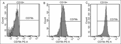 Fig. 1. CD79b expression on B-CLL cells in B-CLL patients, A – negative expression, B – positive CD79b middle expression, C – positive CD79b bright expression.
ZAP-70 expression was determined in 57 patients with Fix&Perm and anti-ZAP-70 antibodies and control, as recommended in literature (17). We also evaluated B-CLL cells count in patients with and without CD79b expression. In both, CD79b-positive and CD79b-negative groups, we evaluated disease stage at diagnosis according to Rai classification, time from diagnosis to the onset of treatment and response to treatment. Patients were monitored for 30 to 48 months.
Additionally, in 50 B-CLL patients with 0/I disease Rai` stage CD100 expression on B-CLL cells was analyzed. In this patient group CD38, CD79b, CD25 and ZAP-70 expression was determined. The control group comprised 10 healthy donors.
Statistical analysis was based on the nonparametric U Mann-Whitney test for independent trials. The p < 0.05 value was accepted as statistically significant. The correlation coefficient was calculated according to Pearson.
Results
CD79b expression on B-CLL cells
CD79b expression on B-CLL cells was detected in 93 patients (82%) while absence of CD79b expression was observed in 20 patients (18%). In the group of CD79b positive patients the mean proportion of CD79b+ B-CLL cells was 78.4 ± 19.7%, (median 83.7). In 80% of CD79b positive patients the value of CD79b+ B-CLL cell percentage was over 60%.
Among CD79b + B-CLL patients we distinguished two groups: one with lower CD79b expression intensity (SI within 3.0-5.9 range) and the other one with higher CD79b expression intensity (SI > 6). Detailed analysis of CD79b expression is presented in table 2.
Table 2. CD79b positive and CD79b negative patients: CD25 and CD38 expression on B-CLL cells.
CD38, ZAP-70 and CD25 expression on B-CLL cells in CD79b positive and CD79b negative patients
CD38 expression was detected in 25% of CD79b positive patients and only in one CD79 negative patient. ZAP-70 expression was determined in 57 patients; 15 patients were ZAP-70 positive (26%). With one exception, all ZAP-70-positive patients belonged to the CD79b+group.
CD25 expression was detected in approximately 90% of CD79b positive and CD79b negative patients. However, the percentage of CD25+ B-CLL cells was significantly higher in CD79b positive patients than that in CD79b negative patients (p = 0.009). The CD25 expression intensity (SI index) on B-CLL cells was also higher in CD79b+ patients than that in CD79b negative patients (p = 0. 05), (tab. 2).
Clinical observations for CD79b-positive and CD79b-negative patients
Studies were performed in the CD79-negative group which included all 20 CLL patients without CD79b expression on B-CLL cells as well as in the group of 30 CLL patients positive toward CD79b antigen, chosen at random. Both groups were identical in terms of gender distribution (proportion of men and women as 1:1). Mean age at diagnosis in the CD79b negative group was 65 ± 9 and 67 ± 12 years in the CD79b-positive group; the difference was not statistically significant (p = 0,56). The observation period for both groups was 30 to 48 months.
Five patients (25%) in the CD79b-negative group did not require treatment, while in 12 patients (55%) we observed disease stabilization with no symptoms of progression. Five patients (20%) progressed and 4 of them died. All patients in the CD79b positive group required treatment. Disease stabilization was observed in 13 patients (43%) while disease progression and/or death in 17 patients (57%). Detailed clinical data of B-CLL course in the studied patients are presented in table 3.
Table 3. Clinical observations of CD79b negative and CD79b positive B- CLL patients.
CD100 expression on B-CLL cells
CD100 expression was determined in 50 patients. The CD100 positive was found in 40/50 patients (80%).
The analysis of the relation between CD100 and CD38 expression shows that in the CD38 positive patient group the percentage of CD100+ B-CLL cells was significantly higher than that in the CD38 negative patient group (respectively: 68.6 ± 16.9 and 33.6 ± 22.6 (p = 0.04). The percentage of CD38+ B-CLL cells in CD100 positive B-CLL patients was higher than in CD100 B negative CLL patients, respectively 15.8 ± 23.8 vs 0.7 ± 0.9 (p = 0.0002). We found a positive correlation between the percentage of CD38+ B-CLL cells and CD100+ B-CLL cells in our patients (r = 0.381, p = 0.007).
Percentage of B CD100+cells in healthy donor peripheral blood was lower than that in B-CLL patients. Contrary to the B-CLL patients, no correlation between analyzed parameters was found in healthy persons. (r = -0.276, p = 0.472). Examples of CD100 expression are presented in figure 2.
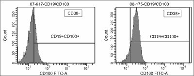 Fig. 2. CD100 expression on B-CLL cells in B-CLL patients: A – CD38 negative patient, B – CD38 positive patient.
In CD100 positive B-CLL patient group noted 36% ZAP-70 positive patients. The percentage CD79b+ B-CLL cells in CD100 positive B-CLL patients was higher than in CD100 B-CLL negative patients: respectively 68.9 ± 31.3 vs 54.9 ± 27.5 (p = 0.1991); CD79b expression intensity (SI) was significantly higher on CD100+ B-CLL cells in positive patients than in CD100 negative patients (p = 0.038).
The percentage of CD25+ B-CLL cells in CD100 B-CLL positive patients was significantly higher than in CD100 negative B-CLL patents (p = 0.0159). Simultaneously, CD25 expression intensity (SI) was higher on CD100 B-CLL cells in positive patients than in negative patients (p = 0.0137). Detailed data are showed in table 4.
Table 4. CD100 expression on B-CLL cells in B-CLL patients.
*non signifcant.
Discussion
Molecular and biological prognostic factors in B-CLL are of significant value due to disease heterogeneity. Apart from such considered molecular factors as chromosomal aberrations (18), immunoglobulin heavy chain gene mutational status, CD38 or ZAP-70 kinase expression (5,19) a search for cytobiological factors determined by flow cytometry methods. CD79b molecule expression is being continued. It has been recently suggested, that a molecule linked with BCR receptor may to be important for prognosis of the course of B-CLL in individual patients (20, 21).
Our clinical observations on CD79b positive patients revealed that they required more often treatment and showed more progressive disease course as compared to CD79b-negative B-CLL patients. Further analysis of the CD79b expression revealed that in patients with CD79b molecule expression on B-CLL cells (especially in those with high intensity), positive ZAP-70 and CD38 expression was more frequent than in patients with low intensity or without of CD79b expression.
This may reflect the relation between disease progression and accumulation of unfavorable prognostic markers in CD79b positive patients. Quijamo et al. (18) showed that markedly high CD79b expression intensity on B-CLL cells is observed in chromosome 12 trisomy cases. Although genetic lesions play a central role in predisposing B-CLL cells to progressive accumulation, they are necessary but not sufficient to maintain the disease. The concept of microenvironment as a regulator of malignant B-cell growth is linked to the possible role of antigen stimulation of BCR signaling (22-25).
Investigation of mechanisms of interaction between the tumor microenvironment and B-CLL cells is an active area of research. CD25, an activating molecule, and CD100 molecule are factors related to interaction of B-CLL cells with the B-CLL cells microenvironment: IL-2 binds CD25 on B-CLL cells and shows an antiapoptotic effect. Exactly our CD79b positive patients showed higher CD25 expression intensity on B-CLL cells than CD79b negative patients.
Nurse like cells (NLC) play an important role in prolongation of B cells survival in B-CLL patients (12, 23). CD100 on B-CLL cell binds plexin on NLC and promotes B-CLL cells survival, parallel to CD38 and CD31 (on NCL cells) interaction (22). In additionally analyzed group 40/50 of patients was CD100 B-CLL positive and showed higher expression of CD25, CD38 and CD79b on CD100+ B-CLL cells than CD100 negative patients.
In our opinion, the estimation of progression risk in B-CLL patients should include not only such known prognostic factors as CD38 and ZAP-70 expression but also the expression of markers for determining the „ability” of B-CLL cells to interact with the B-CLL microenvironment. Apart from molecular studies and detection of already known prognostic factors, B-CLL prognosis should also include broader studies on the immunophenotype of B-CLL cells: CD79b expression as a structure related not only to BCR, but also to CD25, CD38 and CD100 which participate in the interaction between B-CLL cells and B-CLL microenvironment. It is connected with biological diversity of B-CLL and important for prognosis (26, 27).
According to recent reports, detection of high CD79b expression may be also the indication for anti-CD79b monoclonal antibody-based therapy (10, 11).
Conclusions
1. In CD79b positive patients group the „accumulation” of CD38, CD25, and ZAP-70 expression was noted on B-CLL cells and simultaneously the course of disease seems to be worse as compared to CD79b negative group.
2. CD100 positive B-CLL patient` group showed expression of CD38 and CD25 involved, as well as CD100, in interaction between B-CLL cells and the B-CLL microenvironment. Piśmiennictwo
1. Dal-Bo M, Bertoni F, Forconi F et al.: Intrinsic and extrinsic factors influencing the clinical course of B-cell chronic lymphocytic leukemia: prognostic markers with pathogenetic relevance. J Trans Med 2009; 7: 76-90.
2. Gobessi S, Laurenti L, Longo PG et al.: ZAP-70 enhances B-cell-receptor signaling despite absent or inefficient tyrosine kinase activation in chronic lymphocytic leukemia and lymphoma B cells. Blood 2007; 109: 2032-2039.
3. Minuzzo S, Indraccolo S, Tosello V et al.: Heterogeneous intracellular expression of B-cell receptor components in B-cell chronic lymphocytic leukaemia (B-CLL) cells. Br J Haematol 2005; 130: 5504-5509.
4. Treanor B, Depoil D, Gonzalez-Granja A et al.: The membrane skeleton controls diffusion dynamics and signaling through the B receptor. Immunity 2010; 32: 187-199.
5. Boonstra JG, Van Lom K, Langerak AW et al.: CD38 as a prognostic factor in B cell chronic lymphocytic leukemia (B-CLL: Comparison of three approaches to analyze its expression. Cytometry B Clin Cytom 2006; 70: 136-141.
6. Richardson S, Matthews C, Catherwood M et al.: ZAP-70 expression is associated with enhanced ability to respond to migratory and survival signals in B-cell chronic lymphocytic leukemia. Blood 2006; 107: 3584-3592.
7. Deaglio S, Vaisitti T, Bergui I et al.: CD38 i CD100 lead a network of surface receptors relaying positive signals for B-CLL growth and survival. Blood 2005; 105: 3042-3050.
8. Decker T, Bogner C, Oelsner M et al.: Antiapoptotic effect of interleukine-2 (IL-2) in B-CLL cells with low and high affinity IL-2 receptors. Ann Hematol 2010; 89: 1125-1132.
9. Burger JA, Ghia P, Roswald A, Caligaris-Capio F: The microenvironment in mature B-cell malignancies; a target for new treatment strategies. Blood 2009; 114: 3367-3375.
10. Zheng B, Fuji RN, Elkins K et al.: In vivo effects of targeting CD79b with antibodies and antibody-drug conjugates. Mol Cancer Ther 2009; 8: 2937-2946.
11. Dornan D, Bennet F, Chen Y: Therapeutic potential of an anti-CD79b antibody-drug conjugate for the treatment of non-Hodgkin Lymphoma. Blood 2009; 114: 2721-2729.
12. Kurtova A, Balakrishnan K, Chen R et al.: Diverse marrow stromal cells protect CLL cells from spontaneous and drug-induced apoptosis. Blood 2009; 114: 4441-4450.
13. Stevenson FK, Krysov S, Davies AJ et al.: B-cell receptor signaling in chronic lymphocytic leukemia. Blood 2011; 118: 4313-4320.
14. Habib LK, Finn WG: Unsupervised immunophenotypic profiling of chronic lymphocytic leukemia. Cytometry Part B, 2006; 70B: 124-135.
15. Schroers I, Griesinger F, Trumper L et al.: Combined analysis of ZAP-70 and CD38 expression as predictor of disease progression in B-cell chronic lymphocytic leukemia. Leukemia 2005; 19: 750-758.
16. Bigos M, Stovel R, Parks D: Evaluating multicolor fluorescence data quality among different instruments and different laser powers – methods and results. Cytometry 2004; 59A: 42-48.
17. Le Garff-Tavernier M, Ticchioni M, Brissard M et al.: National standardization of ZAP-70 flow cytometry determination: the French experience. Cytometry B Clin Cytom 2007; 72: 103-108.
18. Quijamo C, Lopez A, Rasilio A et al.: Impact of trisomy 12,del(13q0,del(17)p and del(11q) on the immunophenotyp DNA ploidy status and proliferative rate of leukemic-B-cells in chronic lymphocytic leukemia. Cytometry B Clin Cytom 2008; 74: 1441-1445.
19. Kopeć-Szlęzak J. Podstawka U, Sikorska A et al.: Znaczenie CD38 i ZAP-70 jako czynników rokowniczych w przewlekłej białaczce limfocytowej B (PBL-B). Post Nauk Med 2007; 20: 309-314.
20. Van Bockstaele F, Verhasselt B, Philippe J: Prognostic markers in chronic lymphocytic leukemia. Blood Rev 2009; 23: 25-47.
21. Shanafelt TD: Predicting clinical outcome in CLL. Hematology 2009; 75: 421-429.
22. Deaglio S, Malavasi F: Chronic lymphocytic leukemia microenvironment: shifting the balance from apoptpsis to proliferation. Haematologica 2009; 94: 752-756.
23. Patten PE, Buggins AG, Richards J et al.: CD38 expression in chronic lymphocytic leukemia is regulated by the tumor microenvironment. Blood 2008; 111: 5173-5181.
24. Malavasi F, Deaglio S, Damle R et al.: CD38 and chronic lymphocytic leukemia: a decade later. Blood 2011; 118: 3470-3478.
25. Herishanu I, Perez-Galan P, Liu D et al.: The lymph node microenvironment promotes B-cell receptor signaling and tumor proliferation in chronic lymphocytic leukemia. Blood 2011; 117: 563-574.
26. Caligaris- Cappio F.: Inflammation, the microenvironment and chronic lymphocytic leukemia. Haematologica 2011; 96: 353-355.
27. Ching ES, Kumanooh A. Roles of Sema4D and Plexin-B1 in tumor progression. Mol Cancer 2010; 9: 251-260.
otrzymano/received: 2012-05-07 zaakceptowano/accepted: 2012-06-04 Adres/address: *Joanna Kopeć-Szlęzak ul. Bajońska 5/1, 03-963 Warszawa tel.: +48 (22) 617-88-37 e-mail: jszlez@poczta.onet.pl Artykuł Ekspresja molekuł CD79b, CD25 i CD100 na komórkach B w przewlekłej białaczce limfocytowej (PBL-B) w Czytelni Medycznej Borgis. |
  Proszę kliknąć w wybraną okładkę aby przejść na stronę czasopisma
 | |||||||||||||||||||||||||||||||||||||||||||||||||||||||||||||||||||||||||||||||||||||||||||||||||||||||||||||||||||||||||||||||||||||||||||||||||||||||||||||||||||||||||||||||||||||||||||


Chcesz być na bieżąco? Polub nas na Facebooku: strona Wydawnictwa na Facebooku |